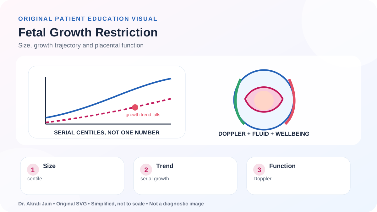
A baby may measure small because they are constitutionally small or because growth is being limited by placental, fetal or maternal factors. Fetal growth restriction (FGR) describes a baby not reaching its growth potential and requires more than a single weight estimate.
RCOG Green-top 31 and 2024 patient guidance
Current guidance separates size from placental function and bases timing of birth on gestation, Doppler findings, growth pattern and maternal condition.
- Risk assessment begins early; high-risk patients may need aspirin and planned serial growth scans.
- Fundal-height measurement from around 24 weeks is a screening tool in lower-risk singleton pregnancy, with ultrasound if growth slows or measurement is small.
- Umbilical-artery Doppler is central when FGR is suspected; very abnormal flow may require specialist fetal-medicine review and earlier birth.
- The latest recommended birth is often near term for an otherwise healthy small baby, while true FGR or maternal disease may justify earlier delivery.
Why babies measure small
Constitutional size may reflect parental stature and remain healthy. FGR is more likely with placental insufficiency, hypertension or pre-eclampsia, smoking, kidney or autoimmune disease, infection, fetal anomaly or genetic conditions.
Accuracy depends on correct pregnancy dating. Ultrasound weight estimates have an inherent margin of error, so trend and additional markers matter.
Ultrasound surveillance
Biometry measures the head, abdomen and femur to estimate weight. Amniotic fluid and placental appearance are reviewed. Umbilical-artery Doppler assesses resistance in placental circulation; other Dopplers may be used in specialist care.
Scans are scheduled at clinically meaningful intervals. Daily scans do not make growth estimates more accurate and can create noise.
Maternal monitoring and movement
Blood pressure and urine are checked because placental dysfunction and pre-eclampsia may coexist. The patient should know their baby’s usual movement pattern and attend promptly for reduction or change.
Bed rest and eating extra calories do not reverse placental FGR. Smoking cessation and treatment of maternal disease are important where relevant.
Timing and mode of birth
The team balances the benefit of more time in the uterus against the risk of deteriorating placental function. Gestation, Doppler results, cardiotocography, growth, fluid and maternal health all influence timing.
Vaginal birth may be possible with reassuring findings and appropriate monitoring. Markedly abnormal Dopplers or fetal compromise may make caesarean birth safer.
Reading the growth picture
Centile
Shows size compared with a reference population, but does not alone prove health or disease.
Trajectory
A falling growth pattern can be more concerning than one isolated measurement.
Doppler and wellbeing
Placental blood-flow and fetal-wellbeing tests help decide surveillance intensity and timing of birth.